

A biomaterial designed to travel through the bloodstream could offer a less invasive way to relieve inflammation and support the self-healing of injured tissue. In animal studies, the injectable material improved tissue damage caused by heart attacks in both rodents and large animals. Initial proof-of-concept experiments also suggested that the same approach could one day be useful for other inflammation-related diseases, including traumatic brain injury and pulmonary arterial hypertension.
“This biomaterial makes it possible to treat damaged tissue from the inside out,” said Karen Christman, professor of bioengineering at the University of California San Diego and lead researcher on the team that developed the material. “It’s a new approach in regenerative medicine.” The results were published in 2022 by a team of bioengineers and physicians in the journal Nature Biomedical Engineering. At the time, Christman explained that a human trial to test the safety and efficacy of the biomaterial could begin within one to two years.
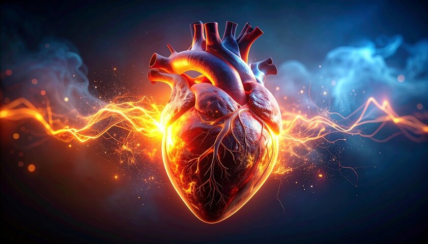
A New Approach to Repairing Heart Damage
Heart attacks remain one of the most serious medical emergencies in the United States, with an estimated 785,000 new cases each year. According to data from the World Health Organization and EU statistics, several million people in Europe also die each year from cardiovascular disease as a whole, and heart attacks and strokes account for a large proportion of these deaths. Cardiovascular diseases account for around a third of all deaths in Europe, making them the most common cause of death.

If the blood flow to the heart is blocked, heart tissue can be damaged or die. The body reacts by forming scar tissue, but this scar does not contract like healthy heart muscle. Over time, this can weaken the heart and contribute to heart failure. There is currently no established therapy that directly repairs heart tissue after a heart attack. Current treatment focuses on restoring blood flow, limiting further damage and managing the risk of future heart problems.
“Coronary artery disease, acute myocardial infarction and heart failure remain the most serious public health problems plaguing our society today,” said Dr. Ryan R. Reeves, a physician in the Department of Cardiovascular Medicine at UC San Diego. “As an interventional cardiologist who treats patients with coronary artery disease and heart failure every day, I would very much like to see another therapy to improve outcomes and alleviate distressing symptoms.”
From Cardiac Hydrogel to Infusion into the Bloodstream
The work builds on previous research by Christman’s team using a hydrogel made from the natural scaffold of heart muscle tissue, also known as extracellular matrix (ECM). This gel was designed so that it could be introduced directly into the damaged heart muscle via a catheter. Once there, it forms a support structure that promotes cell growth and tissue repair. The results of a successful Phase 1 human clinical trial of this earlier hydrogel approach were published in fall 2019. The study found that transendocardial injection of VentriGel, a hydrogel derived from the extracellular matrix of the heart, was safe and feasible in patients following a heart attack with left ventricular dysfunction, although larger randomized trials would be needed to test whether it improves outcomes.
However, the direct injection method has a significant limitation. As it requires an injection into the heart muscle with a needle, it cannot usually be used immediately after a heart attack. Administering it too early could carry the risk of further damage. This challenge led the researchers to another idea: a biomaterial that could be infused into a blood vessel in the heart or administered via an infusion during procedures such as angioplasty or stent implantation.
“We wanted to develop a biomaterial therapy that could be delivered to hard-to-reach organs and tissues, and we found a method that uses the bloodstream – the vessels that already supply blood to these organs and tissues anyway,” said Martin Spang, first author of the study, who is a PhD student in Christman’s group at the Shu Chien-Gene Lay Department of Bioengineering.
Why Intravenous Administration is Important
The bloodstream-based approach gives the biomaterial a major practical advantage. Instead of remaining at a few injection sites, it can be distributed more evenly throughout the damaged tissue. This could make it particularly valuable after a heart attack, when damaged areas may be difficult to reach directly and time is of the essence. The study in Nature Biomedical Engineering described the material as an intravascularly infused extracellular matrix biomaterial made from decellularized, enzymatically digested and fractionated ventricular myocardium. The material was designed to accumulate in the damaged tissue by binding to leaky microvessels and is largely degraded within about three days.

To produce the injectable version, the researchers in Christman’s lab started with the hydrogel they had already developed and tested for compatibility with blood injections. The problem was the particle size. The original hydrogel contained particles that were too large to effectively target damaged, leaking blood vessels.
Spang solved this problem by processing the liquid precursor of the hydrogel in a centrifuge. This allowed the team to separate larger particles and retain only nano-sized particles. The material was then dialyzed, sterile-filtered and freeze-dried. When sterile water is added to the finished powder, the result is a biomaterial that can be administered intravenously or infused into a coronary artery in the heart.
How it Detects Injured Tissue
When the researchers tested the biomaterial in a rodent model of heart attacks, they assumed that it would enter the damaged tissue through leaky blood vessels. After a heart attack, gaps can form between the endothelial cells that line the inside of the blood vessels. Instead, the team observed something more surprising. The biomaterial adhered to these endothelial cells, helping to close the gaps and appearing to accelerate the healing of the blood vessels. This process reduced inflammation, one of the main causes of tissue damage after an injury.
The researchers then tested the treatment on a pig model for heart attacks and achieved similar results. In rats and pigs with induced acute myocardial infarction followed by intracoronary infusion, the biomaterial was associated with reduced left ventricular volume, improved wall motion scores and changes in gene expression associated with tissue repair and inflammation.
Potential Beyond the Heart
Although most of the work focused on heart attack damage, the researchers also tested whether the same biomaterial could be applied to other inflamed tissues. Using rat models, they found proof of concept that the approach could be useful in traumatic brain injury and pulmonary arterial hypertension. This is interesting because both diseases affect organs that are difficult to access: The brain is particularly protected by the blood-brain barrier, while the lungs are very sensitive to inflammation and pressure changes due to their fine vascular network. So if a material acts specifically via the vascular system, it could target precisely those areas where conventional drugs often only have a limited effect.
This broader potential is one of the most fascinating aspects of the work. Many organs and tissues are difficult to access directly, but are all supplied by blood vessels. This is exactly where the idea comes in: The biomaterial not only uses the vessels as a transport route, but actively interacts with their inner wall, the endothelial cells. This means that it could not only work in one organ, but could also be used in various tissues with vascular damage or inflammation – for example after strokes, pneumonia or other forms of acute tissue damage.
If a biomaterial can use these vessels as a “portal of entry”, this opens up new possibilities for regenerative medicine because it can be targeted to where damage occurs without the need for major surgery. “While most of the work in this study focused on the heart, the possibilities for treating other hard-to-access organs and tissues may open up the field of biomaterials and tissue engineering for the treatment of new diseases,” said Spang.
What has Happened Since the 2022 Study
Since the original study, further work has investigated how extracellular matrix-based biomaterials influence regeneration after myocardial infarction. One study published in Nature Communications in 2025 by researchers led by Christman used spatial transcriptomics and single-core RNA sequencing to investigate how injectable extracellular matrix-based biomaterials affect cardiac tissue after myocardial infarction. The study found repair-promoting signals in rat models that included immune modulation, blood vessel and lymphatic vessel development, fibroblast activation, myocardial tissue salvage, smooth muscle cell proliferation and neurogenesis.
While this later work did not replace the need for clinical testing of the intravascular biomaterial, it did provide more detailed insights into how this class of cardiac extracellular matrix therapies can affect healing at the cellular and regional level in damaged hearts. Ventrix Bio, Inc, the start-up co-founded by Christman, has also advanced the development of related cardiac extracellular matrix technology. An entry on ClinicalTrials.gov on VentriGel describes an Emory University-sponsored open-label Phase 1 trial in children with hypoplastic left heart syndrome to evaluate the safety and feasibility of intramyocardial injection of Ventrix Bio’s extracellular matrix material. At the time of access, no participants have been recruited for this study.
Next Steps for Human Testing
Christman and Ventrix Bio plan to apply for FDA approval to study the newer intravascular biomaterial in human heart disease. If approved for clinical trials, the therapy would need to demonstrate that it is safe, convenient to use and effective enough to improve patient outcomes. At present, the treatment is still at the experimental stage. But its appeal is obvious: instead of requiring direct injections into the heart muscle, it could potentially be administered via existing blood vessel-based procedures or intravenously, reaching the damaged tissue from the inside. “A major reason we treat severe coronary artery disease and heart attacks is to prevent left ventricular dysfunction and progression to heart failure,” Dr. Reeves said. “This easy-to-administer therapy has the potential to play a significant role in our treatment approach.”